


Fluorescence spectroscopy of Candida albicans biofilms in bone cavities treated with photodynamic therapy using blue LED (450 nm) and curcumin.
SILVA, Francine Cristina da; RODRIGUES, Paôlla Layanna Fernandes; ARAÚJO, Thalita Santos Dantas; SANTOS, Mariana Sousa; OLIVEIRA, Janeide Muritiba de; ROSA, Luciano Pereira; SANTOS, Gabriel de Pinto de Oliveira; ARAÚJO, Bruno Pereira de; BAGNATO, Vanderlei Salvador.
SILVA, Francine Cristina da; RODRIGUES, Paôlla Layanna Fernandes; ARAÚJO, Thalita Santos Dantas; SANTOS, Mariana Sousa; OLIVEIRA, Janeide Muritiba de; ROSA, Luciano Pereira; SANTOS, Gabriel de Pinto de Oliveira; ARAÚJO, Bruno Pereira de; BAGNATO, Vanderlei Salvador.
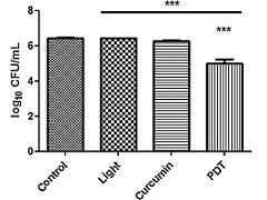 Abstract: Fluorescence spectroscopy may assisst in the diagnosis and control of infectious processes associated with bone lesions of the oral cavity. The aim of this study was to analyze, through fluorescence spectroscopy, Candida albicans biofilms formed in artificial bone cavities treated with photodynamic therapy (PDT) mediated with 450-nm blue light-emitting diode (LED) and curcumin. Another aim of this study was to analyze the existence of a correlation between the effectiveness of the photodynamic treatments and the fluorescence spectroscopy images. Artificial bone lesions (n=40) were made in bovine bones and inoculated with standard suspensions of Candida albicans (ATCC 18804) for biofilm formation (14 days / 36°C ± 1°C). The 40 specimens were distributed among four experimental groups (n=10): L-C- (control), L+C- (LED for 5 min), L-C+ (curcumin for 5 min), and L+C+ (PDT). Aliquots of 100 µL were collected from the bone cavities after treatments and were seeded in duplicate on Sabouraud dextrose agar for 24 h at 36°C ± 1°C and the colony-forming units (CFU/ mL) were counted.Before and after each treatment, the specimens were subjected to spectral fluorescence and the images were compared using the Image J program. The log10 CFU/mL were compared with Kruskal-Wallis and Dunn's Multiple Comparison post-test (significance level at 0.05). The fluorescence histogram values before and after treatment were compared using Wilcoxon test (95%).The correlation between Candida albicans log10 CFU/mL and the number of the fluorescence red pixels spectroscopy was verified using Spearman correlation test. The reduction of Candida albicans log10 CFU/mL in the L+C+ (PDT) group was the most relevant and the fluorescence spectroscopy was correlated to the microbiological result. It was concluded that there was a consistency between the number of Candida albicans log10 CFU/mL and the red pixel data of the fluorescence images, demonstrating that the fluorescence diagnostic device reflects the true microbiological condition of Candida albicans biofilms in the bone cavities during the pre-treatment and post-treatment, thus providing the clinician the ability to dynamically, simply, and instantaneously verify the performance of the treatment used. Abstract: Fluorescence spectroscopy may assisst in the diagnosis and control of infectious processes associated with bone lesions of the oral cavity. The aim of this study was to analyze, through fluorescence spectroscopy, Candida albicans biofilms formed in artificial bone cavities treated with photodynamic therapy (PDT) mediated with 450-nm blue light-emitting diode (LED) and curcumin. Another aim of this study was to analyze the existence of a correlation between the effectiveness of the photodynamic treatments and the fluorescence spectroscopy images. Artificial bone lesions (n=40) were made in bovine bones and inoculated with standard suspensions of Candida albicans (ATCC 18804) for biofilm formation (14 days / 36°C ± 1°C). The 40 specimens were distributed among four experimental groups (n=10): L-C- (control), L+C- (LED for 5 min), L-C+ (curcumin for 5 min), and L+C+ (PDT). Aliquots of 100 µL were collected from the bone cavities after treatments and were seeded in duplicate on Sabouraud dextrose agar for 24 h at 36°C ± 1°C and the colony-forming units (CFU/ mL) were counted.Before and after each treatment, the specimens were subjected to spectral fluorescence and the images were compared using the Image J program. The log10 CFU/mL were compared with Kruskal-Wallis and Dunn's Multiple Comparison post-test (significance level at 0.05). The fluorescence histogram values before and after treatment were compared using Wilcoxon test (95%).The correlation between Candida albicans log10 CFU/mL and the number of the fluorescence red pixels spectroscopy was verified using Spearman correlation test. The reduction of Candida albicans log10 CFU/mL in the L+C+ (PDT) group was the most relevant and the fluorescence spectroscopy was correlated to the microbiological result. It was concluded that there was a consistency between the number of Candida albicans log10 CFU/mL and the red pixel data of the fluorescence images, demonstrating that the fluorescence diagnostic device reflects the true microbiological condition of Candida albicans biofilms in the bone cavities during the pre-treatment and post-treatment, thus providing the clinician the ability to dynamically, simply, and instantaneously verify the performance of the treatment used. | |
| Photodiagnosis and Photodynamic Therapy |
| v. 26, p. 366-370 - Ano: 2019 |
| Fator de Impacto: 2,895 |
| http://dx.doi.org/10.1016/j.pdpdt.2019.05.002 |